

Novel Phantom Characterizes ATCM Performance
/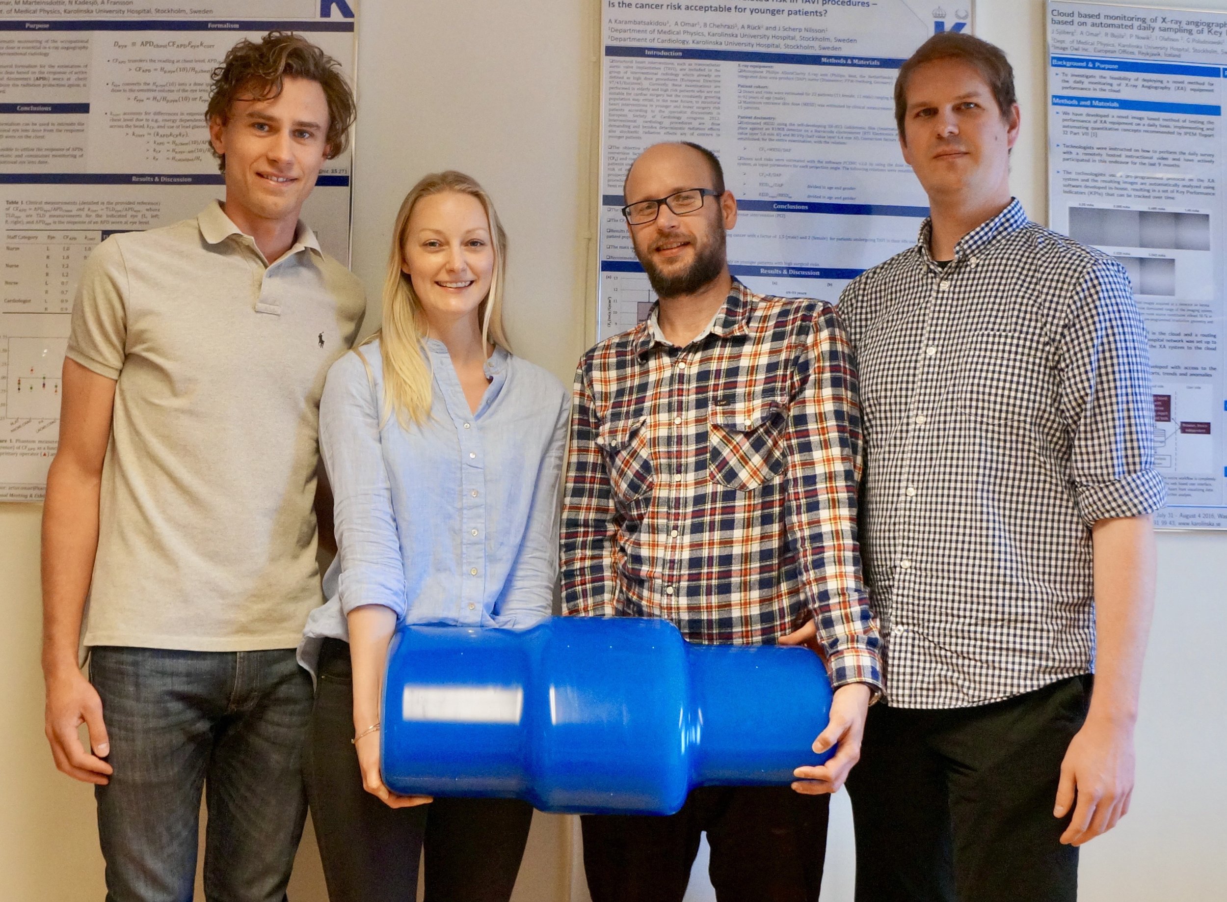
(from left) Patrik Nowik, Deborah Merzan, Gavin Poludniowski, and Robert Bujila with the Karolinska ATCM Phantom
Automatic Tube Current Modulation (ATCM) is an important feature on many modern CT scanners for optimizing patient dose while maintaining acceptable image quality.
The Karolinska University Hospital in Stockholm has a variety of CT scanners from different vendors. Each vendor implements ATCM using its own algorithms. Yet when a team of physicists at the hospital wanted to understand these algorithms better they found their existing phantoms were of limited use. Some investigation revealed that there were no commercially available phantoms that met their requirements to characterize ATCM.

CCT228 ATCM Phantom from The Phantom Laboratory
The team of Deborah Merzan, Patrik Nowik, Gavin Poludniowski, and Robert Bujila decided to take matters into their own hands and design a phantom to allow them to answer the questions they had about how the various ATCM algorithms responded to changes in scanning protocol parameters and body positioning.
The phantom design they settled on features 3 different elliptical cross sections mimicking the attenuation of different sized patients. Special attention was given to creating smooth transitions and eliminating artifacts created by hard edges.
Once the team built their prototype phantom they analyzed scans from a variety of ATCM systems to evaluate image uniformity and noise with different settings and off-center phantom positioning.
The analysis showed significant differences in how the ATCM systems of various CT manufacturers respond to protocol and positioning changes. As team member Robert Bujila remarked, “it is one thing to understand how the ATCM should work in theory (for example by reading the scanner’s documentation) and another thing to understand how the ATCM works in practice...we have found that it is not always straightforward to predict how the ATCM can be affected by variations of the parameters in a scanning protocol.”. Their work provides an important reference on how different ATCM systems function and the evaluation of settings to be considered when adjusting and optimizing scanning protocols [1].
The team presented their work at conferences and ultimately published it in the British Journal of Radiology. The team found that there was widespread interest in the phantom and approached The Phantom Laboratory about commercializing the phantom to make it available for others to use. Bujila said, “We had previously collaborated with The Phantom Laboratory on another project and we felt that it was a natural step to approach [them]”.
Joshua Levy, President of The Phantom Laboratory, was immediately interested. Said Levy, “"The Karolinska imaging physics group has been making important contributions to the imaging field where the balance between image quality and dose are critical. The ACTM phantom they developed is an important tool for the evaluation of Automated Tube Current Modulation. We see this phantom as a natural complement to our Catphan® line of phantoms to characterize the maximum performance of CT scanners”.
The development team at The Phantom Laboratory adapted the prototype design to use the durable, tissue density, Catphan® Uniformity material. Consulting with the Karolinska team at numerous points in the process The Phantom Laboratory has released the phantom as the CT228 ATCM Phantom. Bujila says the team was pleased with the collaboration, “The Phantom Laboratory has been very professional ... We are truly grateful for the dedication and attention to detail that The Phantom Laboratory has had in making our phantom design into a commercial product.”
The Phantom Laboratory is currently working with its long-time software partner Image Owl to automate the analysis of the phantom for Image Owl’s online phantom analysis platform.
The phantom and the automated image analysis will be shown at this year’s AAPM Annual Meeting in Denver, Colorado (The Phantom Laboratory, Booth 5009).
[1]Evaluating the impact of scan settings on automatic tube current modulation in CT using a novel phantom Deborah Merzan, Patrik Nowik, Gavin Poludniowski, and Robert Bujila The British Journal of Radiology 2017 90:1069