Learn about the latest developments in MR QA for radiotherapy
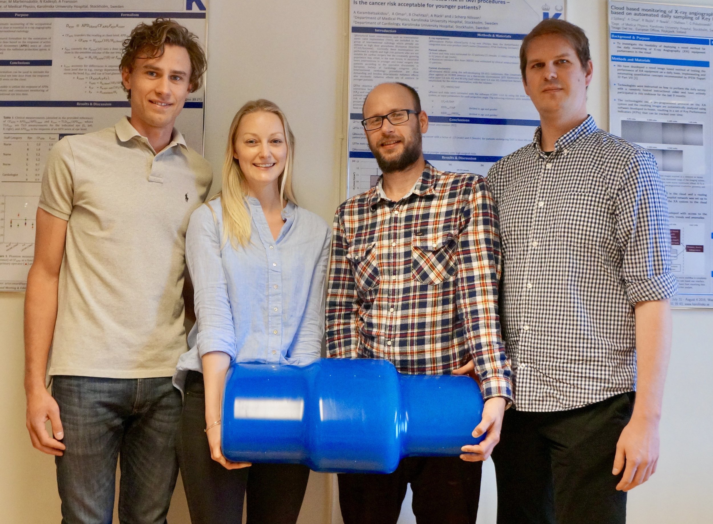
Dr. Richard Mallozzi, MR Physicist and CTO of Image Owl, Inc. will be presenting two 15-minute talks at The Phantom Laboratory's booth (712) during the 2018 AAPM annual meeting in Nashville, TN. on MR QA in radiotherapy.

MR imaging's use for radiotherapy planning and guidance has increased dramatically in the last few years due to its superior imaging of soft tissues for improved image guidance and planning. Adapting MR imaging for use in radiotherapy requires stricter attention to image distortion due to the spatial sensitivity of the applications. However, distortion is not the only imaging parameter that physicists must take into account when evaluating MR images for radiotherapy use.

Dr. Mallozzi will be exploring these issues in 2 talks at various times during the show.

 MRI Quality Control in Radiation Therapy Planning: an Automated Solution
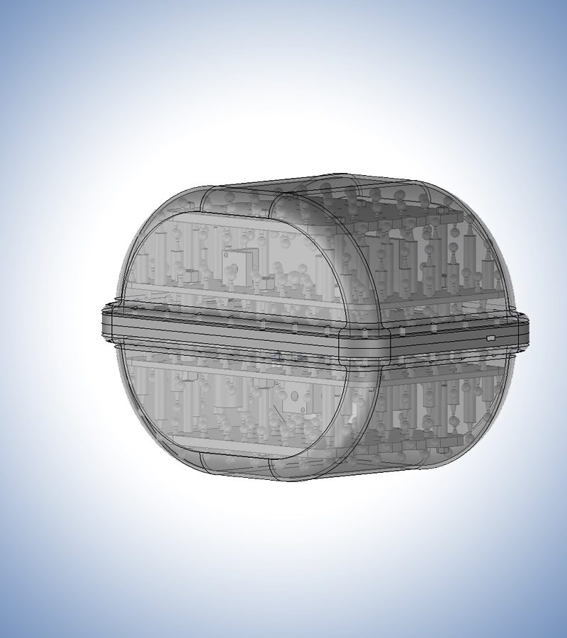
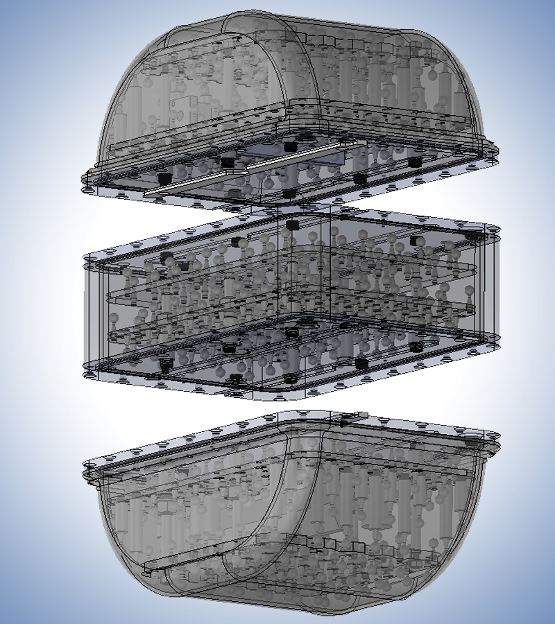
A general discussion of MRI Quality Control for Radiation Therapy Planning, with particular emphasis on geometric distortion. Dr. Mallozzi will present the MagphanRT phantom and its associated Image Owl automated analysis software.

MRI Quality Control in Radiation Therapy Planning: Beyond Distortion
A general discussion of MRI Quality Control for Radiation Therapy Planning, with emphasis on measurements and analysis beyond geometric distortion. Dr. Mallozzi will present the MagphanRT phantom and associated Image Owl automated software analysis of resolution, slice thickness, signal uniformity, signal-to-noise, and laser alignment.

Dr. Richard Mallozzi, MRI Physicist and CTO of Image Owl, Inc.

Richard is a physicist with extensive experience in MRI systems, applications, physics, and engineering. Richard earned an A.B. in Physics from Harvard University, and a Ph.D. in Physics from University of California at Berkeley in 1998, where he studied high-temperature superconductivity. After graduating from U.C. Berkeley, Richard joined GE Global Research as a Magnetic Resonance Scientist, where he worked on diverse projects ranging from MRI acoustic noise reduction, interventional MRI, gradient and RF coil development, and applications of MRI to neuroimaging.

While at GE Global Research, Richard collaborated with the Phantom Laboratory and GE scientist Daniel Blezek to develop the phantom and analysis technique that formed the basis of the control method. Richard joined ONI Medical Systems in 2007, where he helped develop the first high-field (1.5 Tesla) commercial extremity MRI system.

Richard joined Phantom Laboratory and Image Owl in 2014, where he works on a variety of physics and application issues pertinent to medical imaging. he bacame CTO of Image Owl, Inc. in 2017

We look forward to seeing you at AAPM and discussing your needs in this exciting space!


Learn how the most comprehensive phantom for digital breast tomosynthesis was developed.

tomophan 1.jpg

Tomophan® Phantom

Complete Characterization of DBT Image Quality

Digital breast tomosynthesis imaging has become an important technology in mammography screening over the last several years. Its advanced imaging capabilities require specialized tests to fully characterize image quality.

A new paper published in the JACMP describes the design and development of the Tomophan® phantom and its accompanying automated, cloud-based, Tomophan® QA analysis from Image Owl, Inc. 

The development of the phantom follows initiatives from the FDA, the AAPM TG245 task group, and European Reference Organization (EUREF) for Quality Assured Breast
Screening and Diagnostic Services Committee report

Goodenough, D. , Levy, J. , Olafsdottir, H. and Olafsson, I. (2018), Design and development of a phantom for tomosynthesis with potential for automated analysis via the cloud. J Appl Clin Med Phys, 19: 291-300. doi:10.1002/acm2.12297


Looking forward to seeing you at the AAPM Spring Clinical Meeting!

We are looking forward to seeing you at the AAPM Spring Clinical Meeting in Las Vega, April 7-9.  We will be showing our latest developments from The Phantom Laboratory with our partners at Image Owl.

If you are adding MR simulation or guidance to your workflow the Magphan® RT system with automated image analysis tools included provides comprehensive measurement of the key imaging parameters relevant for MR in radiotherapy. Come see the phantom and analysis at table 44  during the spring clinical meeting and discuss your MR QA needs.

UCLA adopts Phantom Laboratory’s Magphan® RT for MR guided radiotherapy QA

Magphan RT 820 phantom with torso coil in the ViewRay MRidian

Magphan RT 820 phantom with torso coil in the ViewRay MRidian

A recent visit by The Phantom Laboratory’s MR physicist Dr. Richard Mallozzi to the UCLA Department of Radiation Oncology helped kick off the use of the Magphan® RT on UCLA’s ViewRay MRIdian MR-guided radiotherapy machine to monitor geometric distortion, uniformity, and other key imaging parameters.

Dr. James Lamb with the Magphan® RT on the ViewRay MRIdian treatment machine.

Dr. James Lamb with the Magphan® RT on the ViewRay MRIdian treatment machine.

UCLA is an early adopter of MR guided radiotherapy. The use of MR guidance in radiotherapy has great benefits in the accurate real-time targeting of tumors with the improved soft tissue contrast of MR imaging. With improved imaging, margins around tumors and organs at risk can often be reduced to less than 3mm compared to 5mm common in conventional IGRT and IMRT treatments. Furthermore, a high proportion of the treatments on the MRIdian machine are abdominal and pelvic SBRTs with 3 to 5 fractions requiring great accuracy.  With these needs for very precise imaging of tumors and organ position, it is critical that MR distortion and uniformity are accurately measured and controlled.

Dr. Mallozzi noted,

“While geometric distortion in modern MR scanners is minimized by the manufacturers, its stability is dependent upon a chain of conditions that should be carefully monitored and controlled.”

Dr. James Lamb, assistant professor and lead physicist for UCLA's ViewRay service, is responsible for implementing the QA procedures for the UCLA’s MRIdian system. He determined early on that daily measurement of MR distortion and uniformity were important as failures in components like coils could be difficult to detect through visual examination of images and could potentially lead to inferior treatments. 

He found that existing QA phantoms for quantifying distortion either failed to cover a sufficient field of view to provide true 3D measurements or were so bulky that they precluded daily use by therapists working on the machine. Furthermore, existing analysis software was not automated so the therapists and physicists could not easily monitor MR image quality

When he learned that The Phantom Laboratory was developing a phantom and analysis to address these shortcomings he was immediately interested.

“We were looking for a comprehensive MR image check”, he commented.
Magphan RT Model 820

Magphan RT Model 820

The Magphan RT phantom’s modular design with all components weighing less than 12 kg makes it practical for a single person to safely set up and handle without special equipment. The design allows the addition of center modules to further extend the field of view or for future additional measurement tools.

The phantom with its attendant analysis service can provide accurate measurements for distortion and uniformity over a 35 x 27 x 21 cm.volume (extendible to 35 x 39 x 21 cm with the addition of a center module). The Magphan RT system can also provide modulation transfer and edge spread functions, slice thickness measurements, signal to noise ratios and positional accuracy measurements. 

The analysis service from Image Owl is hosted online requiring no installation. A simple transfer of the MR DICOM image series starts the fully automated analysis and produces a comprehensive QA report with measurements compared against specifications that provide therapists and physicists a daily snapshot of imaging performance.

Dr. Lamb noted that besides distortion they plan to most closely monitor uniformity and noise as these directly impact targeting quality. In the longer term, they want to track a number of variables to study the stability and characteristics of the guidance MR. As UCLA implements MR planning separate from the ViewRay system, Dr. Lamb plans to use the Maghan RT to characterize and monitor distortion on those scanners as well.

Dr.  Lamb says,

Distortion vs Distance from Isocenter (Part of Magphan RT Report) 

Distortion vs Distance from Isocenter (Part of Magphan RT Report) 

“ With MR guided radiotherapy becoming  more mainstream the Magphan RT fills a significant gap in commercially available phantoms for comprehensive MR imaging QA”

The Magphan RT system will be on display at The Phantom Laboratory/ Image Owl booth #5009 during the AAPM meeting in Denver, CO, July 31 - August 3, 2017

Book an appointment to get a first-hand look at the phantom and analysis service.
 

Novel Phantom Characterizes ATCM Performance

(from left) Patrik Nowik, Deborah Merzan, Gavin Poludniowski, and Robert Bujila with the Karolinska ATCM Phantom

(from left) Patrik Nowik, Deborah Merzan, Gavin Poludniowski, and Robert Bujila with the Karolinska ATCM Phantom

Automatic Tube Current Modulation (ATCM) is an important feature on many modern CT scanners for optimizing patient dose while maintaining acceptable image quality.  
 
The Karolinska University Hospital in Stockholm has a variety of CT scanners from different vendors. Each vendor implements ATCM using its own algorithms.  Yet when a team of physicists at the hospital wanted to understand these algorithms better they found their existing phantoms were of limited use. Some investigation revealed that there were no commercially available phantoms that met their requirements to characterize ATCM.

CCT228 ATCM Phantom from The Phantom Laboratory

CCT228 ATCM Phantom from The Phantom Laboratory

The team of Deborah Merzan, Patrik Nowik, Gavin Poludniowski, and Robert Bujila decided to take matters into their own hands and design a phantom to allow them to answer the questions they had about how the various ATCM algorithms responded to changes in scanning protocol parameters and body positioning.
 
The phantom design they settled on features 3 different elliptical cross sections mimicking the attenuation of different sized patients. Special attention was given to creating smooth transitions and eliminating artifacts created by hard edges.
 
Once the team built their prototype phantom they analyzed scans from a variety of ATCM systems to evaluate image uniformity and noise with different settings and off-center phantom positioning.
 
The analysis showed significant differences in how the ATCM systems of various CT manufacturers respond to protocol and positioning changes. As team member Robert Bujila remarked, “it is one thing to understand how the ATCM should work in theory (for example by reading the scanner’s documentation) and another thing to understand how the ATCM works in practice...we have found that it is not always straightforward to predict how the ATCM can be affected by variations of the parameters in a scanning protocol.”. Their work provides an important reference on how different ATCM systems function and the evaluation of settings to be considered when adjusting and optimizing scanning protocols [1].
 
The team presented their work at conferences and ultimately published it in the British Journal of Radiology. The team found that there was widespread interest in the phantom and approached The Phantom Laboratory about commercializing the phantom to make it available for others to use.  Bujila said, “We had previously collaborated with The Phantom Laboratory on another project and we felt that it was a natural step to approach  [them]”. 
 
Joshua Levy, President of The Phantom Laboratory, was immediately interested. Said Levy, “"The Karolinska imaging physics group has been making important contributions to the imaging field where the balance between image quality and dose are critical. The ACTM phantom they developed is an important tool for the evaluation of Automated Tube Current Modulation. We see this phantom as a natural complement to our Catphan® line of phantoms to characterize the maximum performance of CT scanners”.

The development team at The Phantom Laboratory adapted the prototype design to use the durable, tissue density, Catphan® Uniformity material. Consulting with the Karolinska team at numerous points in the process The Phantom Laboratory has released the phantom as the CT228 ATCM Phantom. Bujila says the team was pleased with the collaboration, “The Phantom Laboratory has been very professional ... We are truly grateful for the dedication and attention to detail that The Phantom Laboratory has had in making our phantom design into a commercial product.”

The Phantom Laboratory is currently working with its long-time software partner Image Owl to automate the analysis of the phantom for Image Owl’s online phantom analysis platform.
 
The phantom and the automated image analysis will be shown at this year’s AAPM Annual Meeting in Denver, Colorado (The Phantom Laboratory, Booth 5009).
 

[1]Evaluating the impact of scan settings on automatic tube current modulation in CT using a novel phantom Deborah Merzan, Patrik Nowik, Gavin Poludniowski, and Robert Bujila The British Journal of Radiology 2017 90:1069

The Phantom Laboratory Attains ISO13485 Certification

FOR IMMEDIATE RELEASE: October 26, 2016

Megan Stalter
The Phantom Laboratory, Inc.
Phone: 518-692-1190

info@phantomlab.com


The Phantom Laboratory Attains ISO13485 Certification

Greenwich, NY:  The Phantom Laboratory announces that it has completed the transition from ISO 9001 to the ISO 13485:2003 standard for the design and manufacture of phantoms for the medical imaging and radiation therapy fields.

The ISO 13485 standard enhances The Phantom Laboratory’s existing quality system in risk management, supplied material traceability, labeling, packaging, sterilization and cleanliness. The enhanced certificate allows Phantom laboratory to successfully meet its customers’ evolving requirements and meet changing regulatory requirements both in the US and internationally.

Josh Levy, Phantom Laboratory’s president said “Our goal is to see our customers use our tools as part of comprehensive quality systems that improve their institutions’ care. In walking our talk, we have expanded our quality system to include ISO13485. This is in line with our commitment to continual improvement. Our work on this was led by Ariel Epstein Dickson, Manager of Quality Systems, and supported by all our employees. I am proud of our team.”

The Phantom Laboratory manufactures dependable, high-precision phantoms and innovative custom solutions for the medical imaging and radiation therapy fields. Founded in 1989, The Phantom Laboratory has been in the forefront of providing tools to medical physicists and imaging scientists to accurately measure the full capabilities of their imaging systems in diverse modalities including CT, MRI, mammography, PET and others.

###

Newly Published: Method and phantom to study combined effects of in-plane (x,y) and z-axis resolution for 3D CT imaging

This newly published paper in the Journal of Applied Clinical Medical Physics by Dr. David Goodenough et al. details the physics behind the Wave module contained in the The Phantom Laboratory's Catphan® 700 phantom and automatically analyzed by the Image Owl Catphan QA service.

The Wave phantom can be used to sample the 3D resolution properties of a CT image, including in–plane (x,y) and z-axis information. The key development in this Wave Phantom is the incorporation of a z-axis aspect of a more traditional step (bar) resolution gauge phantom. 

Magphan® RT: An integrated MR QA system for radiotherapy

The Phantom Laboratory and Image Owl announce the introduction of Magphan® RT for MR QA in radiotherapy. Magphan RT measures the key performance measurements of MR imagers used in radiotherapy planning and guidance. The system consists of a modular, easy-to-handle phantom with automated software to analyze and track performance.

It has become increasingly important to quantify key parameters such as geometric distortion in MR images as they become increasingly used in radiotherapy. The Magphan RT phantom measures distortion with precisely position spheres spaced throughout the phantom volume. Measured and known positions are compared by the analysis software to produce a three dimensional distortion map.

The Magphan RT phantom large size configuration consists of self-contained top and bottom modules that fit together precisely. An optional center module can be placed between the top and bottom modules to produce a super size configuration. This modular design provides measurement over the range of torso sizes commonly encountered in clinical practice while ensuring that the phantom can be handled by a single medical physicist or therapist without special equipment.

The Magphan RT measures a number of important MR image characteristics that can indicate system degradation or failure. These include distortion, uniformity, resolution, slice thickness and signal to noise ratio.

The included analysis service provided by Image Owl’s Total QA® is accessible on any web enabled device and requires no installation. The service analyzes, tracks, presents and stores  all the data associated with Magphan RT.

For more information contact info@phantomlab.com or info@imageowl.com.